| Organism | Microscopy | Culture and identification | Non-culture methods |
| General | Histopath PAS & silver stains etc.
K0H with Parker ink (skin scrapings/nails) |
SAB (Saborauds dextrose agar) medium +/- antibiotics +/- cycloheximide
For other media, see here. Blue thymol blue sticky tape preparation from cultures for examining hyphae and fruiting structures |
MALDI-TOF for ID
Invasive fungal disease: 1-3 Beta-D-Glucan assay (serum) – broad spectrum but not incl. Cryptococcus |
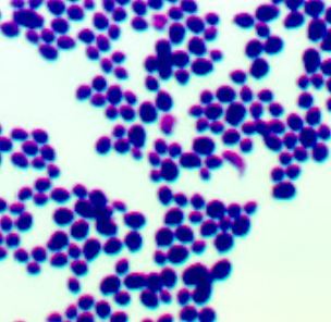
| Candida albicans and other similar species | Characteristic yeast shapes larger than bacteria; appear Gram positive | Colonies with ‘feet’ (C. albicans)
Chromogenic candida agar – usually sufficient for ID. Several types. Yeast sugar assimilation panels e.g. API , VITEK etc |
|
| C. neoformans
C. gattii |
India ink preparation for capsule (CSF)
NB. capsule production media dependent |
Presumptive: capsule+yeast= Cryptococcus (no other species has a capsule)
Urease positive Bird seed agar – pigmented colonies = Cryptococcus CGB medium- (L-Canavanine, glycine, 2 bromthymol blue agar) distinguishes C. gattii from C. neof. |
Crypto antigen lateral flow assay (serum/CSF)
IF serotyping of isolates- research only |
| Aspergillus species | Histopath – narrow septate hyphae; acute angle branching | Culture appearance- pigmentation
Species ID by sticky tape preparation |
Galactomannan EIA (serum, BAL)
Serum precipitins used in ABPA Dx
|
| Pneumocystis jirovecii(PJP) | Bronchial lavage / induced sputum – IF / silver / Giemsa stains | Usually a clinical diagnosis
No culturable
|
PCR more sensitive than IF |
Histopathologic Diagnosis of Fungal Infections in the 21st Century : excellent overview- invasive fungal infections are often recognised only by histopathologists post mortem! Also this slide set : https://www.slideshare.net/appyakshay/fungus-in-histopathology
Cryptococcal serum antigen screening for HIV positive patients– previous posting. This ICT has excellent sensitivity and specificity for crypto antigen detection.
India ink preparation Gram stain of Candida species